Calretinin, also known as CALB2 (Calbindin 2), is a calcium-binding protein found in the central and peripheral nervous systems. It plays an important role in calcium signaling, neuronal excitability, and neuroprotection.
It serves as a biomarker in several research areas, including:
- Neuroscience and neurodevelopmental studies
- Diagnosis of malignant mesothelioma
- Identification of specific interneuron subtypes in the brain
- Tumor classification in oncology
- Parkinson’s and Alzheimer’s disease research
Due to its selective expression in certain cell types, CALB2 detection offers valuable insights into disease mechanisms, tissue-specific expression, and cellular functions.
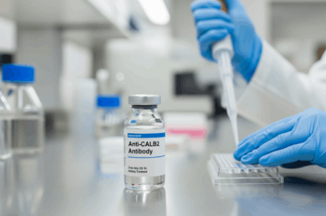
Researchers use anti-CALB2 antibodies in molecular and cellular biology to detect and analyze calretinin protein expression in various biological samples.
So, what are Anti-CALB2 antibodies?
An anti-CALB2 antibody is a type of immunoglobulin that is developed to recognize and bind specifically to calretinin (CALB2) protein. These antibodies can be either monoclonal or polyclonal:
- Monoclonal antibodies are derived from a single B-cell clone. They identify and bind to one epitope on the target protein.
- Polyclonal antibodies are produced by multiple B-cell clones. They recognize and bind to multiple epitopes. So, they provide a stronger signal intensity in some assays.
Depending on the application, researchers can choose an anti-CALB2 antibody type.
What are the Applications of Anti-CALB2 Antibody?
Anti-CALB2 antibody is a cornerstone of various applications, including:
1. Western Blot
Western blotting is an analytical technique used to detect and quantify specific proteins in a sample. In this technique, anti-CALB2 antibodies are used as primary detection agents for calretinin so that researchers can assess protein expression levels in various tissues and cell types.
WB using anti-CALB2 helps identify calretinin-positive interneurons in cortex, hippocampus, and cerebellum in neuroscience, whereas in oncology, it serves as a tumor marker and helps detect calretinin.
To optimize your results with Anti-CALB2 antibody:
- Use a 1:1000–1:2000 dilution of primary antibody.
- Include a positive control lysate (brain tissue lysate).
2. Immunohistochemistry (IHC)
IHC uses Anti-CALB2 antibody to visualize the spatial distribution of calretinin in tissues and provides both qualitative and semi-quantitative data. This method preserves tissue architecture. So, it is a valuable tool, especially in histopathology.
Common tissues analyzed using IHC and Anti-CALB2 are:
- Brain tissues
- Reproductive organs
- Tumor biopsies
To optimize your results with Anti-CALB2 antibody:
- To validate the staining process and accurate results, use both positive and negative control slides.
- In order to detect specific proteins within tissue sections, rely on FFPE (formalin-fixed paraffin-embedded) tissues.
- Check the sample on two different concentrations.
3. ELISA
ELISA stands for Enzyme-Linked Immunosorbent Assay, which is used to detect the amount of calretinin concentration in various biological samples. This technique is known for its high sensitivity.
Anti-CALB2 antibody in ELISA is used as a detection antibody that binds specifically to the calretinin protein. This antibody is used in various ELISA formats, such as sandwich, indirect, and competitive, where it can either act as a capture antibody or a detection antibody.
To optimize your results in ELISA using anti-CALB2 antibody:
- The sample dilution must fall within the detection range of the standard curve.
The Bottom Line
The Anti-CALB2 antibody is a powerful and specific tool for detecting calretinin protein in various biological systems. These antibodies can detect CALB2 expression patterns with precision, no matter whether you are using them for tumor biomarkers, pathway development, or any other experiment. However, make sure you source the antibodies from a reliable source.